
Brain Scans Reveal Brain Changes in Recovered COVID-19 Patients
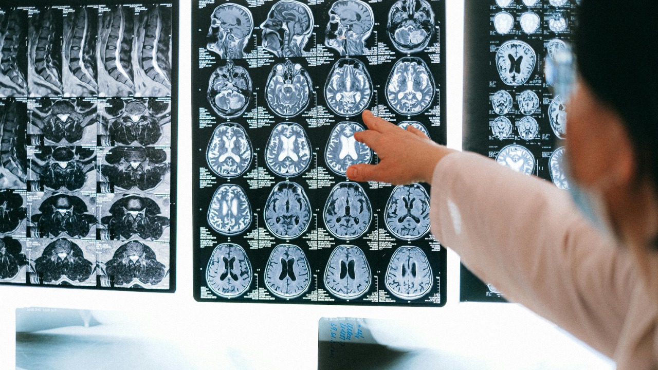
Many individuals who have "recovered" from COVID-19 experience lingering effects such as brain fog: missed appointments, difficulty finding words during conversation, and a mental state that feels muddled. New research using brain imaging indicates these symptoms may be linked to detectable changes in the brain, such as lower metabolic activity and a reduction in gray matter in important areas. In a major study, 785 participants from the UK Biobank were scanned both before and after the pandemic, with 401 who contracted the virus showing distinct structural differences compared to those who remained uninfected.
The Essential Research Supporting the Results
The most closely observed evidence comes from aFlagship Biobank MRI studywhich tracked 785 adults who underwent brain scans prior to the existence of COVID-19, and then again after the virus became widespread. Within this group, 401 individuals tested positive for SARS infections between the two scans and were compared to 384 similar controls who stayed uninfected, with the second scan occurring on average 141 days after diagnosis. Scientists observed a more significant decrease in grey matter thickness and tissue contrast in the orbitofrontal and related areas among those who were infected, suggesting that even mild cases could lead to measurable tissue loss.
These structural results are built upon a more extensive imaging framework that enables this type of research. TheMain Biobank COVID Research Analysis Systemexplains how imaging, biomarkers, medical records, and surveys are combined into one dataset, enabling researchers to compare scans taken before and after an infection, which is uncommon in neurology. The same system also facilitates additional analysis of changes in tissue contrast and other minor indicators that may not be visible on standard clinical scans but could still be connected to thinking abilities and long-term neurological risks.
Brain Changes Patterns Observed
If the Biobank research identifies structural decline, functional imaging demonstrates how the brain functions differently in individuals with long COVID. APrimary PET analysisCompared 35 individuals with long COVID who experienced ongoing neurological symptoms to 44 healthy controls, utilizing voxel-wise methods to analyze metabolism throughout the entire brain. The study identified reduced metabolism in olfactory and orbitofrontal areas, such as the olfactory gyrus, along with involvement of medial temporal structures, extending into the thalamus and brainstem, a pattern that closely matches patients' accounts of smell impairment, memory difficulties, and tiredness.
Further research has aimed to convert that pattern into something healthcare professionals can identify in everyday clinical settings. A multi-centerPrimary PET FDG reference evaluationcentered around a qualitative and consensus-based visual examination of 18F-FDG PET scans in individuals suspected of having neurological long COVID, specifically investigating how frequently the previously identified hypometabolic pattern was observed. By focusing on visual evaluation instead of solely statistical analysis, this research brings the field nearer to developing practical guidelines that nuclear medicine teams can apply when encountering patients who have had COVID and are experiencing unexplained cognitive or sensory issues.
Evidence from MRI and Modern Imaging Techniques
In addition to overall volume reduction, more detailed MRI methods are being explored as possible indicators of long-term effects following COVID-19. APrimary RSNA MRI studyA study prospectively compared 89 patients with Post COVID condition (PCC) to 38 post-COVID controls without impairments, utilizing microstructural MRI features to determine if individual-level classification was feasible. The peer-reviewed research found that specific combinations of microstructural metrics could differentiate PCC from the control group, suggesting that minor white matter or cortical alterations might contribute to ongoing symptoms even when standard MRI scans appear normal.
The seriousness of the initial illness also appears to be significant. In a multi-center high-field imaging study, aPrimary Tesla MRI cohortSeventeen of the 179 participants who were recovered underwent 7 Tesla scans along with comprehensive cognitive assessments. Individuals who had been hospitalized due to COVID-19 exhibited reduced hippocampal volume and lower performance in memory and overall cognitive function tests, such as the MoCA and Trail Making Test, when compared to those who were not hospitalized. One of the primary researchers, mentioned in the PubMed summary, associated these hippocampal changes with the type of forgetfulness and slowed mental processing that many patients report several months after contracting the virus.
What These Modifications Imply for Rehabilitation
Imaging changes are not isolated; they closely align with real-world symptoms like brain fog, attention lapses, and slower processing. A mechanistic study employing dynamic contrast-enhanced MRI revealed that long COVID patients experiencing significant cognitive issues showed signs of blood-brain barrier disruption, with both regional and whole brain leakage measures higher than anticipated. This research, outlined inMain COVID MRI Blood-Brain Barrier studies, also linked the leakage to peripheral blood transcriptomics and in vitro endothelial tests, indicating that vascular and inflammatory mechanisms might contribute to certain cognitive aftereffects.
Functional imaging suggests that some of these abnormalities might show improvement over time, although the pattern of change remains unclear. APrimary US PET FDG COVID Reports groupTracked 45 post-COVID patients who had FDG-PET/CT scans, with 15 of them having pre-infection scans done under the same conditions, and compared these with oncology controls. The researchers noted areas of reduced metabolism in several lobes and the cerebellum that seemed to reach a peak shortly after infection, with almost full recovery seen at later stages, which they viewed as an indication that some of the metabolic issues may be reversible.
Wider Consequences and Their Significance
For medical professionals and decision-makers, these results highlight worries regarding extended neurological risks among a significant number of individuals who have previously been infected. TheBiobank MRI analysisThis found a larger decrease in grey matter thickness and tissue contrast among 401 infected individuals compared to 384 control subjects, indicating that even cases within the community may have minor but detectable brain impacts. When considered alongside microstructural changes in PCC groups and hippocampal volume reduction in more severe instances, it paints a picture of COVID as a condition that can affect various brain systems, not just the lungs.
Radiology groups have begun to present these changes as a justification for organized follow-up instead of a single instance of reassurance. ThePrimary RSNA MRI PCC grouphas been referenced in recommendations for regular cognitive screening and imaging-based monitoring in patients experiencing ongoing symptoms, suggesting that microstructural MRI characteristics might one day direct rehabilitation or drug trials. Similar findings have been reported in young adults, such as a study noted bymental health-related reporting on mild instances of COVID-19, highlights the worry that long-term brain changes are not restricted to older individuals or those who are hospitalized.
Uncertainties and Areas for Future Study
Even with the impressive visuals, scientists are careful about directly linking infection to long-term brain illness. The multi-centerPrimary PET FDG Evaluation for COVID-19Clearly presents its hypometabolic pattern as a correlation rather than evidence of a cause-and-effect relationship, and highlights that differences among individuals and existing health conditions might play a role. Many groups also do not have both pre and post scans for each participant, which makes the Biobank dataset unique and raises uncertainties about how much of the observed changes can be specifically linked to SARS infection versus other stress factors from the pandemic period.
Another unresolved issue is the recovery timeline. While thePrimary US PET FDG COVID Studiescohort indicates almost full restoration of metabolism in numerous patients, other studies, including mechanisticCOVID MRI Blood-Brain Barrier disruption data, highlights continuous leakage and inflammation in individuals experiencing long-term brain fog. Reviews that summarize this developing research, such as those discussing thethe scientific foundation of prolonged COVID-related cognitive difficulties and coverage by European health outlets, emphasize that full reversibility has not been proven yet and that extensive, long-term studies are required.
What Advanced Imaging Is Unveiling Under "Normal" Scans
Several groups are also investigating what occurs in patients whose conventional MRI appears normal. A diffusion microstructure imaging study is described asPrimary DMI MRI COVID PCR testingevaluated hospitalized patients exhibiting neurological symptoms approximately 29 days following a positive PCR test, and observed a change in volume from intra and extra axonal compartments to an increased free water fraction. This pattern aligns with microstructural damage that standard imaging may not detect, suggesting that certain neurological symptoms might indicate minor white matter alterations instead of visible lesions.
Other researchers are connecting these advanced imaging indicators with cellular processes. Reporting on long COVID groups in whichPrimary PET COVID analysisis paired with laboratory assays suggesting that ongoing immune activation and endothelial dysfunction may be key factors contributing to both hypometabolism and BBB leakage. Science communication platforms have emphasized these results as part of a larger initiative to clarify why brain fog can continue even after respiratory symptoms subside, including articles fromscience news sites and general science coveragetaking technical imaging to a broader public.
Expanding the Scope to Prenatal and Child Exposure
Even though most imaging data has primarily focused on adults, similar research is starting to explore the impact of COVID exposure on developing brains. Pediatric studies have found that prenatal infection may be linked to changes in brain structure at birth, as outlined inPrimary prenatal COVID brain imaging coverageFollow-up reports from child health organizations indicate that these early changes in newborns could be linked to future developmental variations, although the research remains preliminary and comes with significant caveats.
Groups focused on population health have also raised issues regarding exposure in the womb and its potential impact on future mental well-being. A summary of findings by academic epidemiologists, presented byinfectious disease researchers, connects prenatal exposure to COVID with brain changes along with developmental delays, anxiety, and depression in young children. Although these results are somewhat separate from adult PET and MRI data, they suggest a similar trend: COVID is not merely a short-term respiratory illness, but a virus that has the ability to alter brain structure and function throughout life, in ways that scientists are just starting to understand.
More from Morning Overview
- 'Failure to shut down': Microsoft issues urgent Windows update warning
- "The road has disappeared": 1,000 people stranded on Outer Banks as highway disappears into the ocean
- Older Tesla vehicles are experiencing issues that owners did not anticipate.
- Apple sends a significant alert to 800 million iPhone users immediately
*This article was researched using AI assistance, with human editors responsible for the final content.
Posting Komentar untuk "Brain Scans Reveal Brain Changes in Recovered COVID-19 Patients"
Please Leave a wise comment, Thank you